By Jessica Lien
A slight change in facial expression, a minimal shift in someone’s tone of voice, or a subtle movement can offer clues about what another person is feeling or thinking, and what they might do next. For many people, interpreting those signals happens almost automatically.
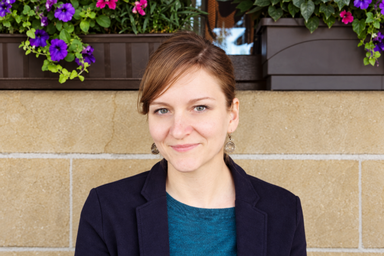
Dorit Kliemann, assistant professor in the Department of Psychological and Brain Sciences, wants to understand how the brain makes that possible.
Kliemann remembers a key moment in her research, when a study participant had entered the lab, and nothing about the interaction prior to the neuroimaging research scan suggested anything unusual. The participant made easy small talk with Kliemann and kindly laughed at a small joke to break the ice.
Then the brain scan appeared on the screen of the console where Kliemann was sitting. Though a typical brain shows two full hemispheres, this one showed a massive gap. The participant, who had undergone a hemispherectomy to treat epilepsy at an earlier age, had nearly half their brain removed. Yet, nothing about Kliemann’s social interaction with the participant suggested the extent of the surgery.
“In some circumstances, the brain is capable of compensating for that kind of massive brain tissue loss, and that’s fascinating,” Kliemann said.
Combining behavioral observations with neuroimaging techniques, Kliemann’s research helps people understand changes in the brain and behavior when people have difficulty with social cognition, as occurs, for example, in autism spectrum disorder or after brain injury.
“I study how our brains help us make sense of other people,” Kliemann said. “I investigate how different parts of the brain work together to support social behavior, and I also am interested in how the brain adapts after substantial injury.”
The fascinating “social brain”
Several conditions can make reading social cues difficult. Kliemann wants to know what separates a brain that reads these cues easily from one that does not: a question she pursues by studying autistic adults and those who have experienced brain damage due to stroke, tumor, or traumatic injury.
Kliemann recently received a significant grant from the National Institutes of Health to study the brain networks’ underlying social behavior. The project focuses on the role of the medial prefrontal cortex (MPFC), a brain region involved in human social cognition. Kliemann and her team use a combination of research involving individuals with brain lesions and different computational approaches to understand the MPFC’s role in the brain networks that support social cognition.
Research and learning at Iowa
The University of Iowa offers an unusual environment for this type of research, combining a strong tradition in psychological theory with cutting-edge cognitive neuroscience. Specifically relevant for Kliemann’s work is the long history of research involving neurological patients via the Iowa Neurological Patient Registry.
“The opportunities for interdisciplinary collaborations here are exciting,” Kliemann said. “I don’t think there is any other place in the world where I could study autism and brain lesions as I am able to do here.”
Trainees are an important part of that work. Depending on their personal and research interests, student trainees in Kliemann’s lab may learn advanced neuroimaging methods, eye tracking, and behavioral research methods. They also learn how to work with autistic adults and people with brain lesions. Kliemann said mentoring also inspires her to reconsider familiar methods and explain complex ideas from new perspectives.
Ultimately, she hopes her work contributes to a deeper understanding of both social behavior and the brain’s capacity to adapt—knowledge that could eventually help clinical researchers better support people with difficulty in social cognition.
“When people learn about my research, I hope they come away with a sense of how remarkable and flexible the human social brain is,” Kliemann said.